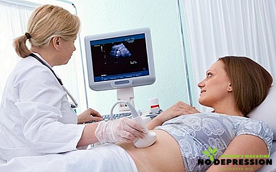
Ultrasound (ultrasound examination) of the urinary system in men and women is a necessary component in the presence of subjective complaints, such as dysuric disorders (frequent, difficult or painful urination), pain, foreign body sensation in the perineum, and the presence of erectile dysfunction in men.
Attention should be paid to changes in laboratory parameters (general analysis of blood, urine in particular) in order to early diagnose diseases and prescribe the necessary treatment.
Ultrasound examination of the prostate in men is a compulsory procedure after 45 years of age and is included in the standard of routine examination (it is possible to diagnose the pathological process in the gland early and start timely treatment).
What does an ultrasound scan of the prostate and urinary organs
In general, the diagnosis of the urinary system allows to evaluate:
- permeability of urine through the ureters;
- the presence of tumor formation in the pelvis and the above-mentioned organs;
- the presence of abnormal suspensions in the cavity of the bladder;
- hypertonus, hypotonia and atony of the bladder, the presence of vesicoureteral reflux;
- the presence of an inflammatory process (acute or chronic);
- presence of diverticula (saccular protrusions of the wall);
- abnormal development of organs (doubling of the kidney or bladder, urethral atresia);
- organ prolapse;
- the presence of tumor formations of the prostate gland;
- condition of the pelvic organs (uterus with its appendages);
To make a correct diagnosis, a more detailed assessment of the organ under investigation with the definition of all its parameters is required.
Ultrasound of the prostate gives the doctor information about the presence of certain diseases of the gland (prostatitis, adenoma, that is, a benign tumor, cancer) by determining its size, clarity of contours, volume, structure homogeneity, the presence of hyperechoic (high density) or hypoechoic (low density) inclusions in gland tissues.
Ultrasound of the urinary system allows you to diagnose the pathology of organs and help the doctor to make the correct diagnosis.
Determined wall thickness, contours, vascular pattern, the size of the urinary system, their position relative to each other (syntopy) and other abdominal organs, the state of the renal pelvis system and the presence of hyperechoic (increased density) formations (stones), the presence of suspensions and pathological formations (diverticula) in the cavity of the bladder.
Also, timely ultrasound diagnostics allows us to determine the congenital malformations of the above-mentioned organs and decide on the possible surgical correction.
How to carry out ultrasound
This table presents various research methods for pathologies of the urinary system and prostate gland:
| Ultrasound Techniques | Prostate | Bladder | Kidney |
|---|---|---|---|
| Transabdominal | + | + | + |
| Transvaginal | + | ||
| Transrectal | + | + | |
| Transurethral | + |
- Transabdominal scan (through the anterior abdominal wall) - a method based on the visualization of organs by external ultrasound. The most common technique and recommended for any age category and any gender.
- Transvaginal scanning, TVUSI (through the walls of the vagina) - a method based on the visualization of the organ (in particular the bladder) in women for a more thorough examination (good visualization due to the absence of subcutaneous fat between the anterior wall of the vagina and the bladder).
- Transrectal scanning, TRUS - a method based on the visualization of organs through the rectum (prostate gland in men; the bladder in women who do not have sex or have contraindications for transabdominal scanning of internal organs).
- Transurethral scanning, TUUSI - a technique used to visualize the urethra and bladder (a connection is established between the pathology of the urethra and the organ described above) using local anesthesia and a contrast agent. Using this method, it is possible to determine the degree of damage to the urethra and the surrounding tissues, to identify possible congenital and acquired defects of its development (agenesis - absence, atresia - underdevelopment, strictures - deformation of its walls).
Ultrasound examination of the bladder with the determination of residual urine
This study has a prognostic value in dysuric disorders (frequent or difficult urination) and indirectly allows the doctor to identify the cause of this condition. Under the residual volume of urine should be understood the amount of fluid remaining in the bladder after urination (should not exceed 50 ml).
The study is carried out in the morning for 3 hours before visiting the toilet (it is important to fix the initial volume of fluid) and after the act of urination (the volume of residual urine should not exceed 10% of the initial).
If the doctor has recorded excess of indicators, it is necessary to conduct a number of additional studies, namely: ultrasound of the bladder with an assessment of its walls (thickness, clarity of contours, presence of impurities and diverticula, developmental abnormalities), ureters and kidneys are evaluated (for vesicoureteral reflux ), Ultrasound of the prostate gland in men (inflammatory and neoplastic processes in the tissues of the gland can serve as a barrier to urine outflow).
Preparation for ultrasound
Men
To properly prepare for ultrasound examination of the bladder and prostate in men, a number of rules must be observed:
- On the eve of the study (about 2-3 days), dieting should be avoided (salty, smoked, spicy foods, alcoholic and carbonated drinks, strong tea and coffee, stimulating intestinal peristalsis, as well as cabbage and legumes - to initiate intestinal loops swelling, which significantly complicates the visualization of organs).
- In the evening before the ultrasound, perform a cleansing enema, take a laxative, or insert a glycerin suppository in order to quickly and sufficiently empty the bowel (especially before the transrectal examination of the prostate gland).
- It is desirable to have a filled bladder before the test (mostly transabdominal), for which you need to either drink a liter of liquid without gas before the test, or not go to the toilet 6 hours before the test (before the transrectal examination of the prostate, filling the bladder is optional and consistent with your doctor).
- If there is a concomitant pathology of the rectum (hemorrhoids, polyps, fissures, tumor processes), you should immediately tell your doctor about this.
It is not recommended to conduct a study in the postoperative period, with the planned preparation for gastroscopy (instrumental examination of the stomach) and colonoscopy (instrumental examination of the large intestine) or in the presence of open wounds on the anterior abdominal wall of the patient (ultrasound examination is postponed for an indefinite period).
It is not recommended to conduct a study in the case of acute respiratory viral infections, acute intestinal infection in a patient (ultrasound is difficult due to adverse patient general health, hyperthermia, pain syndrome).
Women
Preparation for ultrasound of the bladder in women is not significantly different from that in men.
It is necessary:
- Before a transabdominal scan, stick to a diet for several days, have a full bladder for a clearer visualization of the organ.
- Before the transrectal scan, the necessary component is the empty intestine (cleansing enema or laxative in the evening before the test).
- Before transvaginal scanning with additional visualization of the pelvic organs (uterus with appendages), the filled bladder is an optional condition (visualization is not difficult). This access is restricted to non-sexually active persons.
Price of the procedure
- the average price in Moscow and the Moscow region ranges from 800 to 1200 rubles;
- the average price in St. Petersburg is 780-800 rubles;
- The average price in the regions of Russia ranges from 500 to 600 rubles.