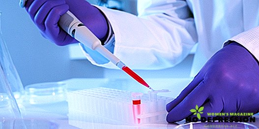
The clinical blood test (AS) is also called full or common. The right to conduct it have a doctor or nurse.
What is a clinical blood test?
According to the general analysis, it is possible to reveal how the blood reacts to the course of various processes and changes within the body. It also allows you to diagnose anemia (low hemoglobin or anemia) and record the beginning and course of development of any inflammatory process.
The study should provide information on the following indicators:
- Erythrocytes (Er, Er).
 This group of cells is called red blood cells. It is one of the most numerous and, above all, provides tissues with oxygen. Also, erythrocyte cells regulate the water-salt balance, transport antibodies and immune complexes, are among the elements that ensure blood clotting.
The erythrocyte is shaped like a biconcave disc and does not have a nucleus. Its small size, shape and plasticity make it possible to pass even along the narrowest and winding capillaries. Any distortion of the original parameters of these cells will be reflected in the results of the study.
Excess Er, called erythrocytosis, may be associated with the influence of psycho-emotional and physical activity. Another pathology variant is an abnormal increase in the size of red blood cells (erythremia) usually due to impaired blood formation. Significant blood loss, hemolysis and anemia can lead to a deficiency of the studied cells, erythropenia.
- Hemoglobin (Hb).
This pigment (dye) contains iron and protein and is a component of red blood cells, providing the possibility of gas exchange in the tissues and the preservation of acid-base balance.
Reducing the number of blood cells with red pigment, respectively, causes a decrease in hemoglobin level, but in some cases there is a significant number of red blood cells that do not contain Hb, that is, the amount of hemoglobin is still insufficient, which allows you to diagnose anemia and prescribe a comprehensive examination of the patient to identify specific sources of the disease .
- Hematocrit.
This indicator reflects the percentage ratio between the fallen Rs and the total blood volume.
- Color indicator (analogue - MCH).
Allows you to assess the level of saturation of erythrocytes with pigment. To identify this indicator, a special formula is used (the ratio of three times the hemoglobin density to the first three digits of the number of red blood cells).
- Erythrocyte volume, mean value (MCV).
Determined by adding the volume of medium, small, large and very large cells and identify their average value. The indicator is significant in the diagnosis of the ratio of water and salt in the body and in the case of identifying the exact type of anemia.
- Leukocytes.
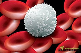

Another name for white blood cells is white blood cells. They do not contain hemoglobin and they are much smaller than red blood cells.
This class of cells is heterogeneous in composition.
The number of leukocytes increases significantly in infectious and inflammatory processes.
- The content and concentration of blood pigment in red blood cells (mean values, MCHC).
For the calculation, hemoglobin and hematocrit level indications are used. Too low result allows you to diagnose hypochromic anemia or thalassemia.
- Erythrocyte anisocytosis (RDW).
Allows you to demonstrate the diversity of red blood cells.
- The rate at which erythrocytes settle (ESR).
An indicator of a non-specific nature, used in identifying the whole list of pathologies of the human body, therefore, it is almost never complete without it. The level of ESR is determined by gender and age.
When conducting UAC, the results of the study of this indicator fit into the lower part of the form and complete the analysis as a whole. Usually, an ESR measurement takes 1 hour.
- Neutrophils.
A group of phagocytic cells that is activated when an infection is ingested.

- Basophils.
An elevated level of basophils indicates an early allergic reaction.
- Eosinophils.
An increase in the number of eosinophils speaks of allergy, worm infestation, or the onset of a recovery phase.
- Lymphocytes.
Provide cellular and humoral immunity. The increased value is recorded at the chronic stage of the disease or if the patient has gone on the mend.
- Platelets.
Increased platelet volume and fluctuations in platelet indices are taken into account when identifying:
- Myeloproliferative disease;
- Infectious inflammatory disease;
- Malignant neoplasms.
In addition, excessive exercise, labor or surgery may affect the increase in this indicator. Platelet count may decrease.
Autoimmune processes, infectious diseases, atherosclerosis, and massive transfusions affect this. A slight decrease in rates observed before the onset of menstruation or during pregnancy.
Indications for use, preparation
The reason for conducting HOW can be almost any disease or prophylactic examination. At the stage of preparation for blood donation, in order not to distort some values, it is recommended to avoid the following factors:
- excessive emotional stress;
- too intense physical exertion;
- food (8-12 hours before the procedure);
- smoking and drinking alcoholic beverages;

- use of a number of drugs;
- too much exposure to ultraviolet radiation (it is better not to sunbathe).

For women, the list of problem points is somewhat expanded:
- passing through the phase of ovulation (the number of leukocytes increases, and on the contrary, there are fewer eosinophils);
- prenatal and birth period (an excess of neutrophils);
- menstruation and pain caused by them (a general distortion of the analysis results).
Ignoring the above points can lead to obtaining biased data and the subsequent lack of proper treatment.
How is a complete blood count?
Sampling of biological fluid in the KLA is done on an empty stomach. Material for the study is taken from the finger (usually using the nameless) or veins, together with the samples for analysis on biochemistry, but distributed into a special tube, in which there is an anticoagulant - EDTA.
In the case of newborns or infants, a special kind of microcontainers is used (also with EDTA). They are suitable for taking material from the finger, heel or earlobe.

Capillary blood gives a slightly different result than venous. In the second method, the indicators of the number of Er and Hb will be much higher, but it is still used more often, because:
- the degree of cell trauma is reduced;
- blood practically does not contact with the skin;
- venous blood is usually taken in an amount sufficient to carry out a repeated analysis on occasion, or to conduct a larger number of studies than was originally intended.
Finally, many people tolerate venous blood sampling much more easily than piercing the skin on a finger.

Results of a clinical blood test: decoding and rate in adults
It should be immediately noted that the concept of the norm in AS is not absolute. Documentation of various medical sources may indicate their own values, but in general they are not very different from those listed here. The inconsistency in the data is registered due to the use of different research methods and analytical systems.
It is much better if the specialist is engaged in deciphering the results, but the patient can also deal with this issue himself, if he understands the meaning of abbreviations and has an idea of what the established standards for each of them are.
| Abbreviation | Decryption | Units | Regulations | |
|---|---|---|---|---|
| Men's | Women's | |||
| RBC | Red blood cell count | 1012 cells per liter | 4,3 - 5,0 | 3,7 - 4,5 |
| HBG, Hb | Hemoglobin | g / l | 129 - 161 | 119 - 141 |
| HCT | Hematocrit | % | 38 - 50 | 34 - 46 |
| ESR | Eir sedimentation rate | mm / h | 1 - 10 | 2 - 15 |
| CPU | Color indicator | - | 0,82 - 1,0 | |
| MCV | RBC Volume (Average) | fl (femtoliter) | 81 - 100 | |
| MCH | Amount of Hb in Ayr (average) | pc (picogram) | 25 - 35 | |
| RET | Reticulocytes (germ Ayr) | Interest (ppm) | 0,21 - 1,21 (2,1 - 12,1) | |
| Mchc | Concentration of Hb in Ayr (average) | g / deciliter | 2,9 - 36,9 | |
| Rdw | Anisocytosis (variety) Ayr | Interest | 11,4 - 14,6 | |
| WBC | White blood cells | 109/liter | 3,9 - 8,9 | |
| BASO | Basophils | Interest 109/liter | up to 1 up to 0,067 | |
| Eo | Eosinophils | Interest 109/liter | 0,49 - 4,9 0,02 - 0,29 | |
| NEUT | Neutrophils | Interest | 46 - 73 | |
| Bandworm | Interest 109/liter | 1 - 6 0,039 - 0,31 | ||
| Segmental | Interest 109/liter | 46 - 68 1,9 - 5,6 | ||
| Lym | Lymphocyte count | Interest 109/liter | 18,9 - 36,9 1,19 - 3,1 | |
| Mon | Monocyte count | Interest 109/liter | 2,9 - 10,9 0,091 - 0,59 | |
| Plt | Platelet count | 109/liter | 179,9 - 319,9 | |
| MPV | Platelet Volume (Average) | fl or micrometers cubic (micron3) | 7 - 10 | |
| PDW | Platelet Diversity | Percentage (%) | 15 - 17 | |
| PCT | Thrombokrit | Percentage (%) | 0,1 - 0,4 | |
All received information is given in a special form, which must be provided to the attending specialist or patient.
Decoding of indicators of the clinical analysis of blood in children: table
From the moment of birth to the completion of puberty (adolescence), children's blood differs markedly in composition and characteristics from that taken from adults. Therefore, for children and young patients have their own standards for each parameter studied.
| Indicator | Age | Norm |
|---|---|---|
| RBC (1012/liter) | Immediately after birth | 4,39 - 6,61 |
| up to 12 months | 3,59 - 4,91 | |
| up to 6 years | 3,49 - 4,51 | |
| under 12 years old | 3,49 - 4,71 | |
| under 16 years old | 3,59 - 5,11 | |
| HBG, Hb (g / l) | Immediately after birth | 139 - 221 |
| up to 12 months | 99 - 141 | |
| up to 6 years | 119 - 146 | |
| under 16 years old | 114 - 149 | |
| RET (‰) | up to 12 months | 2,9 - 14,9 |
| up to 6 years | 2,9 - 11,9 | |
| under 12 years old | 1,9 - 11,9 | |
| under 16 years old | 1,9 - 10,9 | |
| BASO (%) | for any age | up to 1 |
| EO (%) | up to 12 months | 1,9 - 6,9 |
| under 12 years old | 0,9 - 5,9 | |
| after 12 years | 0,9 - 4,9 | |
| NEUT (%) | up to 12 months | 14,9 - 44,9 |
| up to 6 years | 24,9 - 59,9 | |
| under 12 years old | 34,9 - 64,9 | |
| under 16 years old | 39,9 - 64,9 | |
| LYM (%) | up to 12 months | 38 - 72 |
| up to 6 years | 26 - 60 | |
| under 12 years old | 24 - 54 | |
| under 16 years old | 25 - 50 | |
| MON (%) | up to 12 months | 2 - 12 |
| under 16 years old | 2 - 10 | |
| PLT (109/ l) | up to 12 months | 180 - 400 |
| up to 6 years | 180 - 400 | |
| under 12 years old | 160 - 380 | |
| under 16 years old | 160 - 390 | |
| ESR (mm / hour) | up to 1 month | 0 - 2 |
| up to 12 months | 2 - 12 | |
| under 16 years old | 2 - 10 |
Subtleties during pregnancy
It is impossible to deny that pregnancy causes noticeable changes in the female body. All of them are reflected in the results of cancer.
| Indicator | Norm during pregnancy |
|---|---|
| Red blood cells | 3,5 - 5,6 (1012/ l) |
| Reticulocyte | 0.12 - 2.05 (%) - extreme values are allowed only in the absence of any pathologies in the mother's body (usually this indicator only increases slightly) |
| Hemoglobin | from 110 g / l - this is slightly lower than the usual indicator, since during pregnancy there is an increase in the amount of circulating blood, without changing the number of red blood cells |
| White blood cells | 1 trimester: 4.0 - 9.0 (109/ l) 2 trimester: up to 11.0 (109/ l) 3 trimester: up to 15.0 (109/ l) |
| Lymphocytes | 18 - 19% (the lower limit of the normal norm, which contributes to the preservation of the child, not allowing the mother's body to repel it) |
| Myelocytes | 1 - 2% (some increase in comparison with the normal rate due to an excessive amount of granular leukocytes) |
| ESR | up to 45 mm / h (this is the maximum permissible limit, but in general, this indicator can fluctuate with a certain frequency) |
The remaining indicators usually do not change or their changes fit into the normal rate, and iron deficiency, which affects the level of hemoglobin and some other substances, can be compensated for with special vitamins for pregnant women.
Additional information about the clinical analysis of blood from Dr. Komarovsky - in the next video.