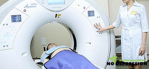
CT of the lungs is a modern diagnostic method that relates to X-ray examinations. This procedure is necessary to study the state and function of the respiratory system, to identify pathological processes. With the help of CT, all segments of the lung can be examined and any changes occurring in the structure of the tissue can be detected.
The essence of computed tomography of the lungs
This method allows to identify the localization of pulmonary tumors and their sizes, determine the stage of development of the pathological process, assess the state of the vessels and lymph nodes, detect changes in the structure of the organ.
The most advanced type of research is spiral CT. Thanks to the latest equipment, you can create detailed images of the studied organ, which are displayed on the monitor screen. Computed tomography is carried out with the introduction of a contrast agent, and without it.
In the first case, before the procedure, the doctor injects a solution based on iodine into the patient's vein. Contrasting is required only in certain cases, for example, to differentiate pathology and better visualization. Computed tomography is characterized by high sensitivity to diseases of the respiratory system.
Indications and contraindications
Carrying out this diagnosis is shown:
- with chest injuries;
- if you suspect the presence of benign or malignant tumors;
- when foreign bodies enter the respiratory organs;
- with an increase in the thoracic lymph nodes;
- if you suspect tuberculosis or other diseases;
- with pathologies of the heart;
- with parasitic lesions of soft tissues;
- pneumonia and pleurisy;
- to detect the source of inflammation and identify the localization of metastases.
The following indications exist for CT using contrasting:
- the need to accurately identify the boundaries of the tumor, to determine the size of the nodes;
- detection of violations of the integrity of blood vessels in the chest;
- assessment of the patient's condition after surgery or his readiness for radiotherapy;
- with suspected pulmonary thromboembolism.
However, this procedure has a number of contraindications, which include:
 severe pathologies of the heart and kidneys;
- diabetes mellitus in the last stage;
- thyroid disease;
- gestation period;
- mental disorders;
- allergic to iodine-containing products, which is important for CT with contrast;
- weight more than 130 kg, since in this case the patient objectively does not have the opportunity to stay in the device for scanning;
- claustrophobia, since the study takes place in confined spaces;
- the overall serious condition of the patient.
 severe pathologies of the heart and kidneys;
In the absence of categorical contraindications, the doctor will set the date of the study.
Features of preparation
Tomography of the lungs does not require a long preliminary preparation, unlike many other diagnostic measures. To prevent serious complications, the doctor may prescribe tests to identify contraindications for CT.
Immediately before the procedure, the patient is required to:
- remove any metal parts and jewelry from the body;
- wear loose clothing;
- 6-7 hours before the scan, stop eating if CT is prescribed with contrast;
- It is necessary to inform the specialist about any chronic diseases and special reactions of the body to any irritants and substances.
The essence of computed tomography of the lungs
CT of the lungs is a modern diagnostic method that relates to X-ray examinations. This procedure is necessary to study the function of the respiratory system, identify pathological processes.

With the help of CT, it is possible to investigate all segments of the lungs and reveal the presence of any changes that occur in the structure of tissues.
Computed tomography with contrast is performed in the same way. The only difference is the preliminary injection of the substance intravenously. Due to this, individual objects in the image of particular interest will be extremely clear. The procedure will take about 30-40 minutes.
CT of the lungs with contrast allows you to easily detect any pathological processes occurring in the pleural region. This procedure is required to diagnose the vessels of the chest area and determine the presence of pathological processes.
What does CT show?
Thanks to computed tomography of the lungs, you can visualize:
- tumors and neoplasms;
- pleural discharge of accumulated fluid inside;
- aortic abnormalities;
- large tuberculous foci;
- bronchitis, bronchopleural fistula.
Computed tomography allows you to assess whether the condition of the lungs is within normal limits. In the pictures, inflammation and any other changes appear as clearly as possible, which makes it possible to make an accurate diagnosis.
The study of the lungs is prescribed as needed, so it is carried out as many times as necessary to make a diagnosis. The rate of radiation load on the body is not exceeded if CT is performed no more than twice a year.
It is possible to carry out this procedure to the child from the first days of life, but only if it is really necessary.
Despite the fact that this diagnostic event is quite safe, during the X-ray radiation is still affected by the organism. That is why children are prescribed it only when other methods of diagnosis do not allow to make a diagnosis.
Benefits of CT
Computed tomography has the following advantages:
- the ability to get detailed pictures that will give the doctor comprehensive information about the state of the lungs;
- short duration of the procedure;
- simplicity and ease of holding;
- efficiency of obtaining the result;
- high accuracy;
- no discomfort during diagnosis.
Decoding results
 To decipher the results of CT does not take much time. After the diagnosis, a specialist examines the resulting images. Each image is evaluated for the density of lung segments, the presence of granulomas in the tissues.
The procedure also makes it possible to determine the boundaries of existing cancer tumors, the presence of pathological foci. The patient will receive CT scan results within two hours after the test.
The cost of CT depends on the city and the status of the clinic where the patient is diagnosed. In Moscow, the price of this procedure varies from 3,000 to 13,000 rubles.
Features of spiral computed tomography
Spiral computed tomography is the most modern type of computerized radiation diagnosis. It is designed to obtain the most accurate images, which is achieved thanks to a thin plane X-ray and computer simulation of the resulting image or video.
The survey is carried out at the expense of special devices and sensors on which the rays fall. Usually, the motion of the tomograph base is complemented by the spiral-shaped rotation of the detector emitters, which makes it possible to form cuts at any given angle.
You can get the clearest picture that gives accurate data thanks to additional detectors about 1 mm thick. At the same time, radiation exposure is even lower than with conventional CT. The study time at MSCT is significantly reduced: the session takes about 20 seconds. Therefore, it is possible to carry out the procedure for seriously ill patients who can not be in a lying position for a long time.

This computer diagnostics can also be supplemented by the introduction of a contrast agent into the vessels. Due to this, the quality of the survey is increased several times.
What are the alternatives?
Computed tomography of the lungs, if necessary, supplemented or replaced by other diagnostic procedures. These include:
- MSCT examination of the lungs. This procedure is carried out with the use of a special drug that reduces the radiation load on the patient. This technique is suitable for detecting tumors, including the esophagus and fistula.
- Coronary angiography. Is an x-ray examination of the coronary vessels using a contrast agent.
- Radiographic computed tomography. One of the most valuable methods of diagnosis, through which you can identify any pathological processes occurring in the area of the lungs or bronchi. If necessary, a patient is injected with a contrast agent, which allows to reliably determine the location of the pathological foci.