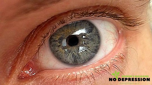
Retinal detachment is often diagnosed already in advanced stages, which complicates treatment. Information about the disease, including the main symptoms, methods of restoring vision, will help to eliminate such situations.
 Human vision is the most complex biological system in which the retina performs one of the most important functions. This fabric is a shell of the eyeball and a kind of conductor. Due to the complex structure (10 layers), including specific receptors, nerve fibers, blood vessels and some other compounds, the retina converts light pulses into nerve ones.
It is they who transmit information to the brain, a person sees, is oriented in space, is able to distinguish objects at different distances and has other abilities characteristic of normal vision.
What is retinal detachment
The retina, including the pigment layer, fits tightly to the choroid, through which the main nourishment of the cells takes place. When an exudate or a special internal fluid from the vessels penetrates between these tissues, then detachment occurs. It can start from the periphery, and only then move to the central part. Formed cavities worsen the supply of nutrients to the layers of the shell, which leads to problems with the perception of the image, in advanced cases - to the death of certain areas.
 Pathology can spread quickly enough, taking measures to prevent detachment is needed as soon as possible so that there is an opportunity to preserve vision.
To provoke such a phenomenon can internal changes in the body, as well as some external influences.
Finding out exactly what caused the disease is an important diagnostic measure, since on the basis of these data a decision is made on the method of treatment.
The reasons
Retinal detachment can occur due to a direct injury when mechanical tissue damage is recorded, or due to internal changes in certain reactions in the body. The reasons may be the following pathological processes:
- diabetes, bleeding disorders;
- oncological conditions;
- infections, including viral type;
- serious diseases of the vascular system;
- various types of fundus lesions, especially concentrated in the central part;
- retinal artery occlusion.
The above diseases, the consequence of which may be retinal detachment, are not the only, but one of the most common. A more accurate cause is determined by the ophthalmologist together with other narrow specialists after diagnostic studies.
In addition to the direct pathologies that can cause dysfunction of the visual system, there are a number of provoking factors. These include:
- pregnancy, difficult or prolonged labor;
- hard work, exercise-related activities;
- genetic predisposition;
- congenital anomalies;
- age changes;
- myopia.
Causes of retinal detachment can be combined, have a different etiology. These features will affect treatment methods as well as the classification of the disease.
Varieties of the disease
To indicate the causes of detachment, the degree of mobility, as well as the area of spread of the affected areas, it is customary to classify tissue damage into separate types. If we consider the disease in relation to provoking factors, the following categories are distinguished:
- Regmatogenous (primary) detachment. Formation of thinning layers due to
 violation of metabolic reactions aimed at nourishing cells. The process occurs due to vascular insufficiency. In the absence of measures to normalize the structure of blood vessels, detachment occurs deeper, extends to other areas of the retina.
 - Exudative (secondary). The development is provoked by infection of the surrounding tissues or the retina itself, the pathologies transferred as well as oncological formations.
- Traction. Diagnosed infrequently, characterized by a strong tension of the retina relative to the vitreous body, which leads to damage.
- Traumatic. This type of detachment may appear immediately after injury or after some time.
In addition to the classification regarding the causal circumstances of the disease, there is a division according to the extent of damage prevalence. The following types are distinguished by these parameters:
- Local Diagnosed with retinal lesions of no more than 25%, is considered the initial degree of development of damage.
- Common If tissues have changed by more than 50%, then detachment is considered to be already common.
- Subtotal. With lesions within 75%, the vision is in critical condition, the high risk of its complete loss.
- Total. Retinal detachment is fixed over the entire surface of the eye, visual function is completely lost, recovery is impossible.
In addition to the listed types of damage to the shell, there are differences in the degree of mobility - rigid or mobile.
The main symptoms
The process of detachment occurs gradually, accompanied by certain signs. At the initial stage, they are short-lived and dimly pronounced, but over time the situation worsens, the symptoms become especially noticeable.
In the initial stages, the following symptoms are characteristic:
- Specific "photo effects". It seems that there was a flash, lightning, an abrupt change in photosensitivity. All signs are short-lived, after a while the normal perception of the image is restored.
- The appearance of flies, points, thin strings. The extraneous forms appear already at the developing detachment, the frequency varies depending on the degree and depth of the tissue damage.
- Loss of clarity, blurriness, the appearance of the shroud. Signs indicate significant retinal lesions, and immediate medical attention is required.

It is impossible to skip the symptoms of retinal detachment, but the main thing is to consult an ophthalmologist in time, even if vision problems are temporary.
Diagnostics
If the ophthalmologist on symptomatology and visual examination suspects retinal pathology, then the patient is sent for a comprehensive examination. All activities are aimed at studying the state of the retina, the possible causes of damage to the eye membrane.
For each case is determined by its list of diagnostic procedures, but when considering the standard range of diagnostic measures can be distinguished:
- Measurement of intraocular pressure (tonometry), affecting the state of not only the visual system, but also the organism as a whole.
- Visual acuity, or viziometriya. Information is necessary for the analysis of measurements of acuity, if it is sharply reduced, this signals the development of pathological processes.
- Exploring the possibility of visual field, or perimetry. In the case of reduced boundaries, the diagnosis of retinal detachment is considered confirmed;
- Structural examination of the eye (biomicroscopy).
- Analysis of the general state of the fundus.
In addition to ophthalmologic studies, special diagnostic procedures may be prescribed to clarify the disease, factors that trigger structural changes in the eyeball. Ultrasound examinations, analyzes of biological material, including blood, urine, as well as electrooculography, electroretinography are practiced.

Treatment methods for retinal detachment
It is possible to restore or remove the affected areas only by surgery, the principle of which is chosen individually in each case. The goal of any intervention is the normalization of cell nutrition, the elimination of injuries, breaks, and the stimulation of the closest fit of the layers.
This is achieved by the following methods:
- Laser exposure. The areas are treated with a laser beam, cauterized, which leads to the limitation of the further propagation of damage on the surface of the membrane, enhances the connection between the pigment and vascular tissues. It is used for minor defects. The operation may have a different cost depending on the size of the damaged area, the complexity of the manipulations. The price varies from 11,000 to 27,000.
- Extrascleral methods. Performed outside or on the surface of the sclera. Distinguish between filling, when special connecting elements (seals) strengthen and bring together the layers, as well as ballooning. The latter option involves the introduction of a balloon with a catheter, the creation of a certain pressure on the tissue, the subsequent fixation of the membrane by laser irradiation. Both procedures require special professionalism from the doctor, the selection of technology is carried out with respect to the identified damage to the eye. The cost depends on the affected area, ranges from 30,000 to 115,000.
- Endovitreal manipulation, or vitrectomy. The intervention involves the correction or removal of the vitreous body, the operation takes place from the inside. The retina is spliced, including laser, with an artificial substitute for the vitreous body, previously treated with special solutions. It is practiced in relatively advanced cases, when the causes of visual impairment are structural changes in the eyeball. The operation requires special skills from the surgeon, the cost is not fixed, since much depends on the complexity, the average price is from 27,000 to 89,000 rubles.
Folk remedies
Despite the existence of prescriptions for treating a disease, the proven fact is that it is impossible to eliminate the tissue defect that has appeared. Attempts to get rid of the problem on their own can lead to a worsening of the situation, the formation of other pathologies of the visual system. The best option is not to experiment, when symptoms appear, immediately consult an ophthalmologist.
Prevention
To prevent retinal detachment, you should periodically visit a doctor to check your eyesight, general eye condition. This is especially true for age patients who have suffered head and eye injuries and have serious chronic diseases.
If possible, you should regulate physical activity, a constant voltage above the norm negatively affects not only the visual function, but also the organism as a whole.
The earlier the retinal detachment is diagnosed, the more gentle the treatment program will be, the more likely it is to maintain the normal functions of the eyes. When running stages there is no guarantee of full return of view, but from a financial point of view, it will require significant costs.


 violation of metabolic reactions aimed at nourishing cells. The process occurs due to vascular insufficiency. In the absence of measures to normalize the structure of blood vessels, detachment occurs deeper, extends to other areas of the retina.


