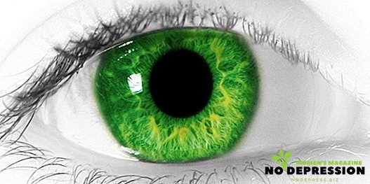
The eyes are a complex body as they contain various work systems that perform many functions aimed at collecting information and transforming it.

The visual system as a whole, including the eyes and all their biological components, includes more than 2 million component units, including retina, lens, cornea, nerves, capillaries and vessels, iris, macula, and optic nerve.
It is imperative for a person to know how to prevent diseases related to ophthalmology in order to maintain visual acuity throughout life.
The structure of the human eye: photo / scheme / drawing with a description

In order to understand what constitutes the human eye, it is best to compare the organ with the camera. Anatomical structure is presented:
- Pupil;
- Cornea (no color, transparent part of the eye);
- Iris (it determines the visual color of the eyes);
- The lens (responsible for visual acuity);
- Ciliary body;
- Retina
The following structures of the eye apparatus also help to ensure vision:
- Vascular membrane;
- Optic nerve;
- The blood supply is made with the help of nerves and capillaries;
- Motor functions are performed by the eye muscles;
- Sclera;
- Vitreous humor (main defense system).

Accordingly, such elements as the cornea, lens and pupil act as the "lens". Light or sunlight falling on them is refracted, then focused on the retina.
The lens is an "autofocus", since its main function is to change the curvature, so that visual acuity is maintained on the norm indicators - the eyes are able to clearly see the surrounding objects at different distances.
The retina works as a kind of “film”. On it remains the seen image, which is then in the form of signals, transmitted through the optic nerve to the brain, where the processing and analysis takes place.

To know the general features of the structure of the human eye is necessary to understand the principles of work, methods of prevention and treatment of diseases. It is no secret that the human body and each of its organs is constantly being improved, which is why, in evolutionary terms, the eyes managed to achieve a complex structure.
Due to this, various structures of biology are closely interconnected - vessels, capillaries and nerves, pigment cells, connective tissue actively participates in the structure of the eye. All these elements help the coordinated work of the organ of vision.
Anatomy of the structure of the eye: the main structures
The eyeball, or directly the human eye, is round. It is located in the deepening of the skull, called the orbit. This is necessary because the eye is a delicate structure that is very easily damaged.
The protective function is performed by the upper and lower eyelids. The visual movement of the eyes is provided by the external muscles, which are called oculomotor muscles.
The eyes need constant hydration - this is the function of the lacrimal glands. The film formed by them additionally protects the eyes. The glands also provide an outflow of tears.

Another structure related to the structure of the eyes and ensuring their direct function is the outer shell - the conjunctiva. It is also located on the inner surface of the upper and lower eyelids, is thin and transparent. The function is gliding during eye movement and blinking.
The anatomical structure of the human eye is such that it has another, more important for the organ of vision, the sclera. It is located on the front surface, almost in the center of the organ of vision (eyeball). The color of this formation is completely transparent, the structure is convex.
Directly transparent part is called the cornea. That it has a heightened sensitivity to various kinds of irritants. This happens due to the presence of a multitude of nerve endings in the cornea. The absence of pigmentation (transparency) allows the light to penetrate inside.

The next eye membrane that forms this important organ is vascular. In addition to providing the eyes with the necessary amount of blood, this element is also responsible for the regulation of tone. The structure is located inside the sclera, lining it.
Each person's eyes have a certain color. For this feature is responsible structure, called the iris. Differences in shades are due to the content of the pigment in the very first (outer) layer.
That is why the eye color is not the same for different people. The pupil is a hole in the center of the iris. Through it, the light penetrates directly into each eye.
The retina, despite being the thinnest structure, is the most important structure for quality and visual acuity. At its core, the retina is a nerve tissue composed of several layers.

The main optic nerve is formed from this element. That is why visual acuity, the presence of various defects in the form of hyperopia or myopia is determined by the state of the retina.
Vitreous body called the cavity of the eye. It is transparent, soft, almost jelly-like. The main function of education is to maintain and fix the retina in the position necessary for its work.
Optical system of the eye
The eyes are one of the most anatomically complex organs. They are the "window" through which a person sees everything that surrounds him. This function allows you to perform an optical system consisting of several complex, interrelated structures. The structure of "eye optics" includes:
- Cornea;
- The lens;
- Retina.
Accordingly, the visual functions they perform are light transmission, refraction, and perception. It is important to remember that the degree of transparency depends on the state of all these elements, therefore, for example, if the lens is damaged, a person begins to see the picture clearly, as if in a haze.
The main element of refraction is the cornea. The luminous flux enters it first, and only then enters the pupil. It, in turn, is the diaphragm on which the light additionally refracts, focuses. As a result, the eye receives an image with high definition and detail.

Additionally, the function of refraction and produces the lens. After a luminous flux hits it, the lens processes it, then transfers it further to the retina. Here the image is "imprinted".
The fluid and the vitreous are a little refractive. However, the state of these structures, their transparency, a sufficient amount, have a great influence on the quality of a person’s vision.
The normal operation of the ophthalmic optical system leads to the fact that the light falling on it passes the refraction, processing. As a result, the image on the retina is reduced in size, but completely identical with the real ones.
Also note that it is upside down. A person sees the objects correctly, since the finally “printed” information is processed in the corresponding sections of the brain. That is why all the elements of the eyes, including the vessels, are closely interrelated. Any slight violation of them leads to loss of sharpness and quality of vision.
The principle of the human eye
Based on the functions of each of the anatomical structures, you can compare the principle of the eye with the camera. The light or image passes first through the pupil, then penetrates the lens, and from there into the retina, where it is focused and processed.
Disruption of their work leads to color blindness. After the refraction of the light flux, the retina translates the information imprinted on it into nerve impulses. They then enter the brain, which processes it and displays the final image, which the person sees.

Prevention of eye diseases
Eye health must be constantly maintained at a high level. That is why the issue of prevention is extremely important for any person. Checking visual acuity in a medical office is not the only concern for the eyes.
It is important to monitor the health of the circulatory system, as it ensures the functioning of all systems. Many of the violations identified are due to lack of blood or irregularities in the delivery process.
Nerves - elements that are also important. Damage to them leads to a violation of the quality of vision, for example, the inability to distinguish the details of an object or small elements. That is why you can not overtax your eyes.

With long-term work, it is important to give them rest every 15-30 minutes. Special gymnastics is recommended for those who are involved in work, which is based on long-term consideration of small objects.
In the process of prevention, special attention should be paid to the illumination of the working space. Feeding the body with vitamins and minerals, the consumption of fruits and vegetables helps to prevent many eye diseases.
Do not allow the formation of inflammation, as this can cause suppuration, so proper eye hygiene is a good preventive measure.
Thus, the eyes - a complex object that allows you to see the world around. It is required to take care, to protect them from diseases, then the vision will retain its sharpness for a long period.
The structure of the eye is shown in great detail and clearly in the following video.


